兔/鼠通用型Streptavidin-HRP试剂盒(DAB)

有货

库存信息

关闭
货号 (SKU) 包装规格 是否现货 价格 数量
S665512-3ml
3ml 现货 Stock Image

基本描述

英文名称 SP Rabbit & Mouse HRP Kit(DAB)
储存温度 2-8°C储存
运输条件 冰袋运输
产品介绍

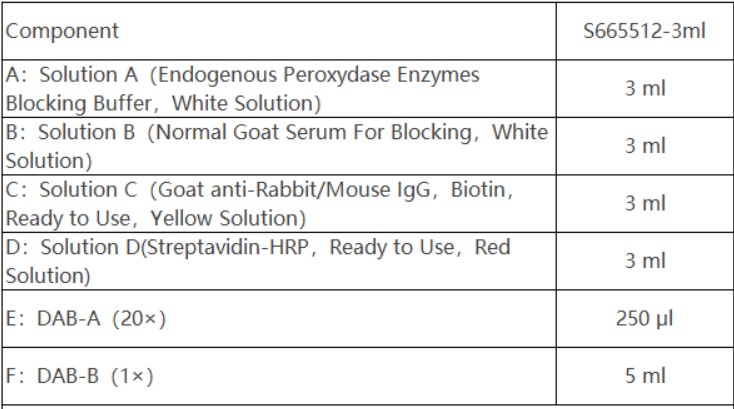
本试剂盒根据生物素(Biotin)与链霉亲和素(Streptavidin)具有很强亲和力的原理设 计。在兔源或鼠源的一抗与相应的靶抗原结合后,本试剂盒中的生物素化抗••兔/鼠通 用型二抗与一抗特异结合;二抗上标记的生物素与标记了过氧化物酶(HRP)的链霉 亲和素结合,形成抗原~特异性一抗~生物素化的二抗~HRP标记的链霉亲和素复合 物。HRP可以催化底物显色,从而推断待检抗原的存在及分布。该试剂盒使用的生物 素化二抗、SA-HRP,均采用优化的标记和纯化技术,使得其染色有更高灵敏度和更低 的背景,适合于检测福尔马林固定石蜡包埋组织切片,以及冰冻切片、细胞爬片、新 鲜制备的血涂片等。兔/鼠通用型Streptavidin-HRP试剂盒适合与我司即用型或浓 缩型抗体配套使用。 

组分:

注意:本试剂盒仅适用于一抗为免源或鼠源抗体的IHC实验

注意事项:

1.以每张切片加入1滴(约50 μl)计算,3ml可做60张切片, 18ml可做360张切片。 

2.对于内源性生物素含量比较丰富的组织,使用本试剂盒时最好用内源性生物素阻断 剂进行封闭。 

3.DAB工作液现配现用,配制好的工作液2-8°C避光1小时内有效。 

4.实验过程中避免组织片干燥,因此各步孵育时工作液用量必需充足,保证完全覆盖 组织样本,且尽量在湿盒中进行孵育。 

5.为获得最佳实验结果,请务必优化实验条件及试剂用量。 

6.DAB为可疑致癌物,使用时请采取必要的防护措施。 

7.本产品仅用于科研,不能用于人体反应或人体治疗

操作步骤 

1.常规处理欲检测的石蜡或冰冻组织切片或细胞爬片等样本。 

1)组织切片或细胞爬片染色前处理: a. 石蜡切片 脱蜡水化:60ºC烤片1小时,二甲苯脱蜡二次,每次5分钟;再依次浸入梯度乙醇 (无水乙醇-无水乙醇-95%-85%-75%乙醇)和蒸馏水中各5分钟水化。 b. 冰冻切片和细胞爬片 切片(或爬片)浸于0.01 M pH7.4 PBS洗涤3次×5分钟。然后用0.1%Triton X-100覆 盖组织(或细胞)浸润15分钟,0.01 M pH7.4 PBS洗涤2次×5分钟。 

2)石蜡切片的抗原修复:绝大大多数情况下,石蜡组织切片用柠檬酸缓冲液高压修 复都是适合的。 修复工作液配制:1 L去离子水中加入10 ml柠檬酸缓冲液(IHC抗原修复液, 100×) ,混匀即可。 修复过程:修复液加入高压锅内,待修复切片浸于修复液中(必需没过组织),盖 上压力锅盖,加热至均匀喷汽,从喷汽开始计时,1~2分钟后压力锅离开热源,自 然冷却至室温,取出切片,蒸馏水淋洗后,再用PBS(0.01 M pH7.4)漂洗2次,每 次3分钟。 

2.滴加适量Solution A白色溶液,即内源性过氧化物酶封闭液室温孵育10分钟,PBS充 分淋洗。 

3.滴加适量Solution B白色溶液,即封闭用正常羊血清工作液,室温孵育10分钟,甩干。 

4.滴加适量一抗工作液(商品化即用型抗体或适当比例稀释的浓缩抗体),按实验要 求孵育,然后PBS充分淋洗。 

5.滴加适量Solution C黄色溶液,即生物素标记羊抗兔/鼠二抗工作液,室温孵育10分钟, PBS充分淋洗。 

6.滴加适量Solution D红色溶液,即HRP标记的链霉亲和素,室温孵育10分钟,PBS 充分淋洗。 

7.DAB显色工作液配制:根据需要量,将DAB-A和DAB-B以1:19的体积比混匀后即 为DAB显色工作液。也可选择每毫升试剂B中滴加1滴(约50 μl)试剂A,混匀即可。 

8.显色:加适量的DAB显色工作液于需要显色的组织切片或细胞爬片上即可显色,显 色时间一般为1-5分钟。显微镜下观察控制显色时间,当达到最佳显色效果后,自来 水冲洗终止显色。显色后的切片经复染、脱水透明,封片后可长期保存。

This reagent kit is designed based on the principle that biotin and Streptavidin have a strong affinity. After the primary antibody of rabbit or mouse origin binds to the corresponding target antigen, the biotinylated antibody in this kit • • Rabbit/mouse universal secondary antibody specifically binds to the primary antibody; The biotin labeled on the secondary antibody binds to streptavidin labeled with peroxidase (HRP), forming an antigen-specific primary antibody biotinylated secondary antibody streptavidin complex labeled with HRP. HRP can catalyze substrate colorimetry, thereby inferring the presence and distribution of the tested antigen. The biotinylated secondary antibody and SA-HRP used in this reagent kit all adopt optimized labeling and purification techniques, which make their staining more sensitive and have a lower background. They are suitable for detecting formalin fixed paraffin embedded tissue sections, as well as frozen sections, cell slides, freshly prepared blood smears, etc. The rabbit/mouse universal Streptavidin HRP kit is suitable for use with aladdin ready to use or concentrated antibodies.

Composition:

Note: This reagent kit is only suitable for IHC experiments where the primary antibody is an immune or mouse derived antibod

Notes:
1. Add 1 drop (approximately 50) to each slice μ l) Calculation: 3ml can make 60 slices, and 18ml can make 360 slices.
2.For tissues with abundant endogenous biotin content, it is best to use endogenous biotin blockers for blocking when using this kit.
3. DAB working solution is prepared and used immediately, and the prepared working solution is effective within 1 hour in the dark at 2-8 ° C.
4. During the experiment, avoid drying the tissue slices, so the amount of working fluid used during each incubation step must be sufficient to ensure complete coverage of the tissue sample, and incubation should be carried out in a wet box as much as possible.
5. To obtain the best experimental results, please make sure to optimize the experimental conditions and reagent dosage.
6. DAB is a suspected carcinogen, please take necessary protective measures when using it. 

7. This product is only for scientific research and cannot be used for human reactions or treatments.

Operation steps:
1. Routine processing of samples such as paraffin or frozen tissue sections or cell slides to be tested.
1) Preparation for staining of tissue sections or cell slides: a. Dewaxing and hydration of paraffin sections: bake at 60 º C for 1 hour, dewaxing twice with xylene for 5 minutes each time; Then immerse in gradient ethanol (anhydrous ethanol anhydrous ethanol 95% 85% 75% ethanol) and distilled water for 5 minutes each for hydration. b. Frozen sections and cell climbing sections (or climbing sections) were soaked in 0.01 M pH 7.4 PBS and washed 3 times for 5 minutes. Then cover the tissue (or cells) with 0.1% Triton X-100 and infiltrate for 15 minutes. Wash twice with 0.01 M pH 7.4 PBS for 5 minutes.
2) Antigen repair of paraffin sections: In most cases, high-pressure repair with citric acid buffer is suitable for paraffin tissue sections. Preparation of repair solution: Add 10 ml of citric acid buffer (IHC antigen repair solution, 100 x) to 1 L of deionized water, and mix well. Repair process: The repair solution is added to a high-pressure cooker, and the repaired slices are immersed in the repair solution (must have no tissue). Cover the pressure cooker cover, heat until evenly sprayed with steam, and start timing from the spraying. After 1-2 minutes, the pressure cooker leaves the heat source and cools naturally to room temperature. Remove the slices, rinse with distilled water, and rinse twice with PBS (0.01 M pH 7.4) for 3 minutes each time.
2. Add an appropriate amount of Solution A white solution, which is an endogenous peroxidase blocking solution, and incubate at room temperature for 10 minutes, then rinse thoroughly with PBS.
3. Add an appropriate amount of Solution B white solution dropwise, which is sealed with normal sheep serum working solution. Incubate at room temperature for 10 minutes and shake dry.
4. Add an appropriate amount of primary antibody working solution (commercial ready to use antibodies or concentrated antibodies diluted in appropriate proportions) dropwise, incubate according to experimental requirements, and then rinse thoroughly with PBS.
5. Add an appropriate amount of Solution C yellow solution, namely biotin labeled sheep anti rabbit/mouse secondary antibody working solution, incubate at room temperature for 10 minutes, and rinse thoroughly with PBS.
6. Add an appropriate amount of Solution D red solution, which is HRP labeled streptavidin. Incubate at room temperature for 10 minutes and rinse thoroughly with PBS.
7. Preparation of DAB color working solution: According to the required amount, mix DAB-A and DAB-B in a volume ratio of 1:19 to obtain DAB color working solution. Alternatively, one drop (approximately 50) can be added per milliliter of reagent B μ l) Reagent A, mix well.
8. Color development: Add an appropriate amount of DAB color development working solution to the tissue section or cell slide that needs to be developed, and the color development time is generally 1-5 minutes. Observe and control the color development time under a microscope. When the optimal color development effect is achieved, rinse with tap water to terminate the color development. The colored slices are re stained, dehydrated and transparent, and can be stored for a long time after sealing.

AI解读

安全和危险性(GHS)

质检证书(CoA,COO,BSE/TSE 和分析图谱)

C of A & Other Certificates(BSE/TSE, COO):
输入批号以搜索分析图谱:

通过匹配包装上的批号来查找并下载产品的 COA,每批产品都进行了严格的验证,您可放心使用!

找到1个结果

批号(Lot Number) 证书类型 日期 货号
G2426657 分析证书 25-05-09 S665512

相关技术文章

FAQ
抗PD-L1的自动化免疫组化染色方案
蛋白质印迹通用实验方案
General Protocol for Western Blotting
蛋白质印迹的裂解物制备法
Methods of Lysate Preparation for Western Blotting
蓝色天然聚丙烯酰胺凝胶电泳实验方案 (BN-PAGE)
BN-PAGE Protocol
组蛋白的蛋白质印迹法
Histone Western Blot
蛋白质去磷酸化实验方案
Protein Dephosphorylation Protocol

溶液计算器